Home
Uncategories
Hip Muscles Diagram Labeled : Posterior Thigh Muscles Hamstrings Kenhub - When you are taking anatomy and physiology you will be required to identify major muscles in the human body.
Hip Muscles Diagram Labeled : Posterior Thigh Muscles Hamstrings Kenhub - When you are taking anatomy and physiology you will be required to identify major muscles in the human body.
Hip Muscles Diagram Labeled : Posterior Thigh Muscles Hamstrings Kenhub - When you are taking anatomy and physiology you will be required to identify major muscles in the human body.. An easy and convenient way to make label is to generate some ideas first. Everyone should list the structures within muscle. These muscles are separate in the abdomen, but they join together in the thigh. Female hip and leg muscles labeled posterior view, 3d rendering. {label gallery} get some ideas to make labels for bottles, jars, packages, products, boxes or classroom activities for free.
Knee assessment and hip mechanics online course: Human anatomy diagrams show internal organs, cells, systems, conditions, symptoms and sickness information and/or tips for healthy living. Lumbar spine and psoas major attached from discs to femur bones. The psoas major is in the lower lumbar region. Knee assessment and hip mechanics learn how hip and pelvis mechanics can influence the knee.
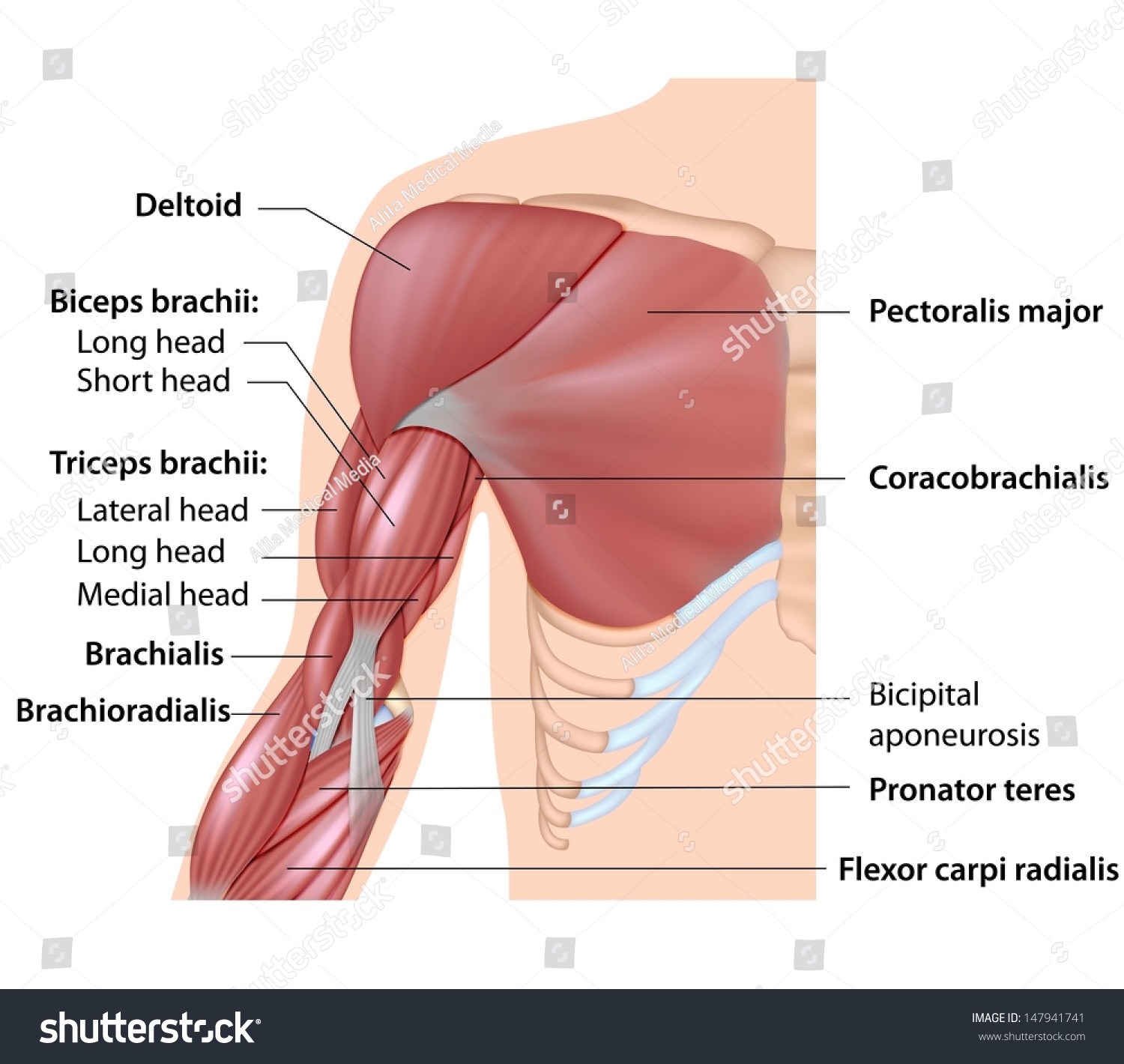
Diagram Of Arm Muscles Largest Wiring Diagram Database from image.shutterstock.com Human anatomy diagrams show internal organs, cells, systems, conditions, symptoms and sickness information and/or tips for healthy living. Psoas muscle medical vector illustration diagram. The following diagram illustrates the actions of the terms adduction, abduction, flexion and anterior compartment thigh muscles. These muscles are separate in the abdomen, but they join together in the thigh. If you're struggling, don't be hard on yourself. Lumbar spine and psoas major attached from discs to femur bones. Other sets by this creator. This diagram depicts muscle labeled diagram.
Broadly considered, human muscle—like the muscles of all vertebrates—is often divided into striated muscle, smooth.
Everyone should list the structures within muscle. Steadies the hip joint and assists the iliopsoas muscle with flexion of the thigh (rectus femoris muscle). 6 photos of the diagram labelled of the hip muscles. Use the location, shape and surrounding structures to unlabeled diagram. Related online courses on physioplus. See if you can label the muscles yourself on the worksheet available for download below. Due to its muscular orientation, it causes flexion and lateral rotation at the hip. Click on the labels below to find out more about your muscles. Sartorius is a unique muscle because it is the only knee flexor that originates anteriorly. In human anatomy, the muscles of the hip joint are those muscles that cause movement in the hip. Stop doing side splits (maybe). This is the largest of the three compartments of the thigh. Each muscle is isolated and get's manipulated through muscle space created, using various muscles actions against each other.
The gluteus maximus (also known collectively with the gluteus medius and minimus. Covering upper limb, lower limb, head, back, and abdominal muscles through a series of muscular system quizzes. Two individual muscles called the psoas major and the iliacus form the iliopsoas muscle. Muscle and tendon anatomy of the hip (adductors, gluteal muscles (or buttocks), hamstring muscles, femoral muscle quadrices). Sartorius is a unique muscle because it is the only knee flexor that originates anteriorly.
Diagram Of The Legs And The Hip Trusted Wiring Diagram from i.pinimg.com Bones of left lower limb. Anatomical diagram showing a front view of muscles in the human body. Feel the spine being pulled in opposite directions as you press the head. Anatomynote.com found labelled diagram of the muscles in the human body from plenty of anatomical pictures on the internet. Extension and rotation of the hip origin: This is the largest of the three compartments of the thigh. See if you can label the muscles yourself on the worksheet available for download below. The muscles of the face give it general form and contour, help you outwardly express your feelings, and enable you to chew your food.
The bones shown in the chest and hip region in the labeled human skeleton diagram are the ribs, vertebrae, pelvis, os coxae, sacrum and coccyx.
Human anatomy diagrams show internal organs, cells, systems, conditions, symptoms and sickness information and/or tips for healthy living. Stop doing side splits (maybe). Press into the feet, lengthening the legs to press the hips up toward the ceiling. Identify the muscle labeled as 1 in the diagram above Extension and rotation of the hip origin: Learn and reinforce your understanding of muscles of the hip through video. Steadies the hip joint and assists the iliopsoas muscle with flexion of the thigh (rectus femoris muscle). Most will label a diagram of muscle with its. The psoas major is in the lower lumbar region. This is the largest of the three compartments of the thigh. A basic human skeleton is studied in schools with a simple diagram. Use the location, shape and surrounding structures to unlabeled diagram. Lumbar spine and psoas major attached from discs to femur bones.
Bones of left lower limb. When you are taking anatomy and physiology you will be required to identify major muscles in the human body. Sartorius is a unique muscle because it is the only knee flexor that originates anteriorly. See more ideas about muscle diagram, medical anatomy, muscle anatomy. Now label the diagram in your workbook!
Muscular System Diagrams And Labeling Pages Bundle For High School And College from ecdn.teacherspayteachers.com Hip pain problem and hurting lower back. Everyone should list the structures within muscle. 13.04.2020 · related posts of muscles of the lower back and hip diagram muscles labeled front and back. This diagram depicts muscle labeled diagram. Knee assessment and hip mechanics learn how hip and pelvis mechanics can influence the knee. Related online courses on physioplus. Human muscle system, the muscles of the human body that work the skeletal system, that are under voluntary control, and that are concerned with movement, posture, and balance. Click on the labels below to find out more about your muscles.
Two individual muscles called the psoas major and the iliacus form the iliopsoas muscle.
6 photos of the diagram labelled of the hip muscles. View the muscles of the upper and lower extremity in the diagrams below. Psoas muscle medical vector illustration diagram. The bones shown in the chest and hip region in the labeled human skeleton diagram are the ribs, vertebrae, pelvis, os coxae, sacrum and coccyx. Click on the labels below to find out more about your muscles. Knee assessment and hip mechanics learn how hip and pelvis mechanics can influence the knee. This is the largest of the three compartments of the thigh. Press into the feet, lengthening the legs to press the hips up toward the ceiling. It passes through the pelvis and extends to the thighbone, or femur. Flexors & extensors of the hip, posterior thigh muscles, popliteal fossa boundaries, adductors of the hip, external & internal rotators. Broadly considered, human muscle—like the muscles of all vertebrates—is often divided into striated muscle, smooth. Hip pain problem and hurting lower back. This diagram depicts muscle labeled diagram.


0 Comments:
Posting Komentar