Home
Uncategories
Blank Leg Bone Diagram : Bones of the Foot Quiz Anatomy : The foot bones shown in this diagram are the talus, navicular, cuneiform, cuboid, metatarsals and calcaneus.
Blank Leg Bone Diagram : Bones of the Foot Quiz Anatomy : The foot bones shown in this diagram are the talus, navicular, cuneiform, cuboid, metatarsals and calcaneus.
Blank Leg Bone Diagram : Bones of the Foot Quiz Anatomy : The foot bones shown in this diagram are the talus, navicular, cuneiform, cuboid, metatarsals and calcaneus.. Blank on the inside, each card is. The tarsal bones and the five long metatarsal bones together form the arches of the foot. Rethinking pain education learn how to teach your patient about their pain powered by physiopedia. Each circuit displays a distinctive voltage condition. Skeletal system diagram without labels printable human skeleton.
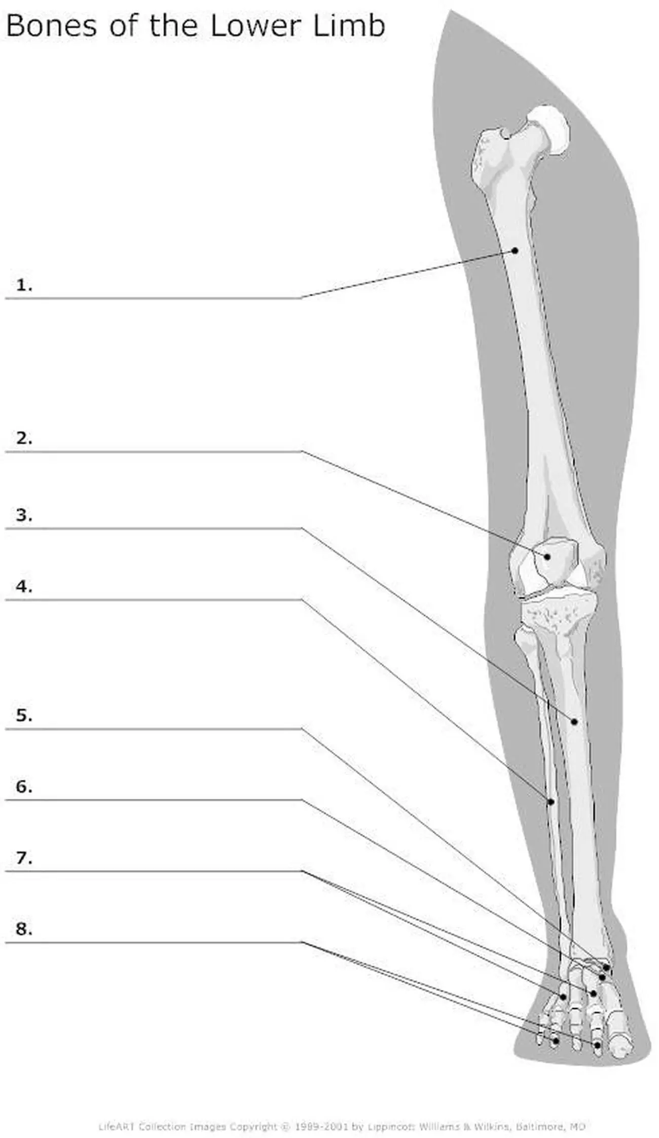
Your leg bones are the longest and strongest bones in your body. Learn vocabulary, terms and more with flashcards, games and other study tools. A fish bone diagram is a common tool used for a cause and effect analysis, where you try to identify possible causes for a certain problem or event. Learn how to draw the femur, patella, tibia, and fibula in this lesson! The tibia is the main bone of the leg, forming what is more commonly known as the shin.
An Introduction to Skeletal System - The Bones and What ... from www.exploringnature.org Learn vocabulary, terms and more with flashcards, games and other study tools. The causes are divided into six main branches in manufacturing, which are collectively referred to as the 6 ms of manufacturing. Learn how to draw the femur, patella, tibia, and fibula in this lesson! Time to jump right into the biggest and strongest bones in the human body. The lungs are found in the thoracic cavity, and extend laterally into the right and left halves around the heart. Master leg and knee anatomy using our topic page. Your leg bones are the longest and strongest bones in your body. Want to learn more about bones?
The rib cage is a structure of bones that surrounds and protects the thoracic cavity, with 12 ribs protecting each of the two lungs.
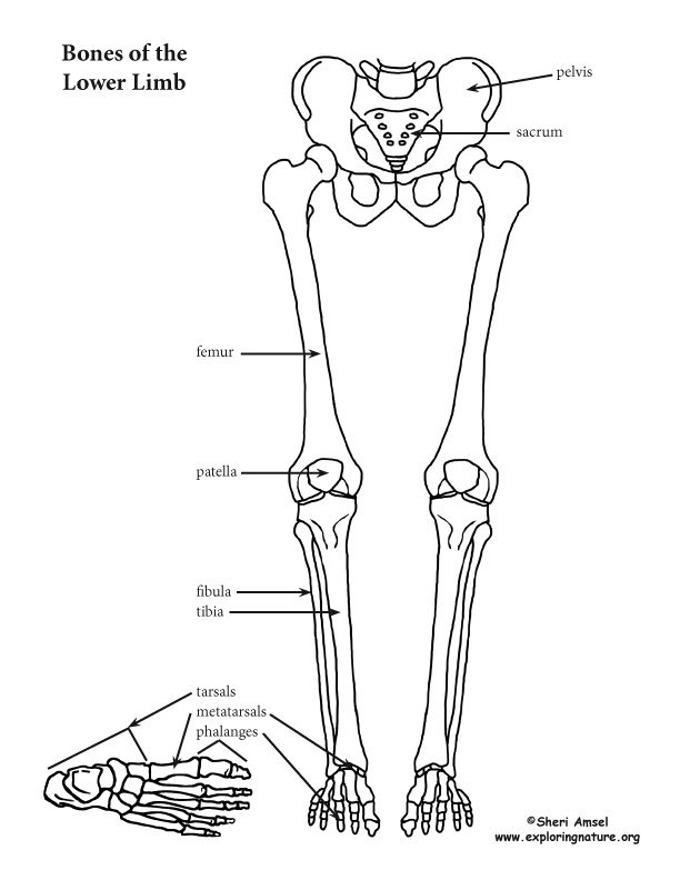
The leg skeleton | anatomy bones, human body anatomy, leg. The tarsal bones and the five long metatarsal bones together form the arches of the foot. Human skeleton, the internal skeleton that serves as a framework for the body. The femur, or thighbone, is the longest and largest bone in the human body. Most bones of the limbs contain mainly yellow bone marrow composed for the most part of fat. This framework consists of many individual bones and cartilages. Learn how to draw the femur, patella, tibia, and fibula in this lesson! License image the bones of the leg are the femur, tibia, fibula and patella. Blank bone diagram creative images. When your muscles contract, they pull the bone they're attached to, making your leg move. A leg bone is a bone found in the leg. Physical performance conflict, for example, difficulties walking or climbing stairs, not being able to keep up, a poor performance in sports (having lost a game, being put on the. Master leg and knee anatomy using our topic page.
The bones mentioned in each human skeleton chart are: 5.3 use the fishbone diagram in sales process. Rethinking pain education learn how to teach your patient about their pain powered by physiopedia. Printable skeletal diagram wiring diagrams click. After that, you need to give bones to the fish.
{Lower Leg Bones Pelvic-Femur-Tibia-Fibula-Foot} | John ... from www.johnthebodyman.com The leg skeleton | anatomy bones, human body anatomy, leg. The foot bones shown in this diagram are the talus, navicular, cuneiform, cuboid, metatarsals and calcaneus. Here's a diagram with the tibia bone labelled, as well as the fibula, showcasing all their surface landmarks. 5.3 use the fishbone diagram in sales process. Lower leg muscle diagram blank sketch coloring page. 467 x 624 jpeg 45 кб. Want to learn more about bones? The bones of the leg are the femur, tibia, fibula and patella.
Want to learn more about bones?
Master leg and knee anatomy using our topic page. Human skeleton, the internal skeleton that serves as a framework for the body. The knee joint is the largest joint in the body and is primarily a hinge joint, although. Printable skeletal diagram wiring diagrams click. Physical performance conflict, for example, difficulties walking or climbing stairs, not being able to keep up, a poor performance in sports (having lost a game, being put on the. The bones of your leg have roughened patches on their surfaces where muscles are attached. Rethinking pain education online course: The foot bones shown in this diagram are the talus, navicular, cuneiform, cuboid, metatarsals and calcaneus. Here's a diagram with the tibia bone labelled, as well as the fibula, showcasing all their surface landmarks. The tarsal bones and the five long metatarsal bones together form the arches of the foot. Rethinking pain education learn how to teach your patient about their pain powered by physiopedia. The following downloads may help you get started, and if you continue reading, i've included some detailed information about how to use the diagrams. 5.3 use the fishbone diagram in sales process.
After that, you need to give bones to the fish. A fish bone diagram is a common tool used for a cause and effect analysis, where you try to identify possible causes for a certain problem or event. Lower jaw (mandible) collar bone. 467 x 624 jpeg 45 кб. The following downloads may help you get started, and if you continue reading, i've included some detailed information about how to use the diagrams.
Pictures Of Bones Of The Lower Extremities from healthiack.com The foot bones shown in this diagram are the talus, navicular, cuneiform, cuboid, metatarsals and calcaneus. Master leg and knee anatomy using our topic page. Skull, clavicle, mandible, scapula, thorax, sternum, humerus, ulna, radius, carpus, phalanges (fingers), metacarpus, spine, pelvis, sacrum, femur, tibia, fibula, tarsus. When your muscles contract, they pull the bone they're attached to, making your leg move. The foot bones shown in this diagram are the talus, navicular, cuneiform, cuboid, metatarsals and calcaneus. Learn vocabulary, terms and more with flashcards, games and other study tools. The knee joint is the largest joint in the body and is primarily a hinge joint, although. This article has clear diagrams/pictoral representations which i would like to use for teaching purposes.
Lower jaw (mandible) collar bone.
Each circuit displays a distinctive voltage condition. Want to learn more about bones? The lungs are found in the thoracic cavity, and extend laterally into the right and left halves around the heart. The leg skeleton | anatomy bones, human body anatomy, leg. The foot bones shown in this diagram are the talus, navicular, cuneiform, cuboid, metatarsals and calcaneus. Editor · aug 13, 2017 ·. Lower jaw (mandible) collar bone. The foot bones shown in this diagram are the talus, navicular, cuneiform, cuboid, metatarsals and calcaneus. Learn more about the leg and knee anatomy by taking our special quiz, customized to focus on bones, muscles, nerves and vessels of. Rethinking pain education online course: Printable skeletal diagram wiring diagrams click. This bright and colorful worksheet helps your child bring the technical terms of the bones in his legs down to size. Cheek bone (zygoma) upper jaw (maxilla).
Bone marrow is the soft, highly vascular and flexible connective tissue within bone cavities which serve as the primary site of new blood cell production or hematopoiesis leg bone diagram. Your leg bones are the longest and strongest bones in your body.


0 Comments:
Posting Komentar